 |
 |
||
|
Characterisation of the Physical, Chemical and Electronic Properties of Solids on the Nanometre Scale |
|||
|
|
||
|
Characterisation of the Physical, Chemical and Electronic Properties of Solids on the Nanometre Scale |
|||
Positron Re-emission Microscopy (PRM) The contrast mechanism for PRM lies in the sensitivity of positron re-emission probability to both surface conditions, including ie the presence of surface defects, adsorbed molecules, different crystal faces, local charging, and to trapping centres in the bulk, predominantly vacancy-type defects. In the only other working PRM positrons are focused into a microbeam which impinges on to the back face of a thin Ni foil. A fraction of the positrons emerge from the other side of the foil and an image is obtained of the exit surface; images are obtained of the Ni itself or of any large adsorbate structures deposited on the film. |
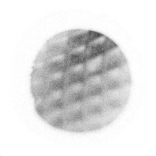 An image of a nickel mesh over a tungsten backing taken with the microscope
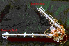
The electrostaticlenses of the microscope |
|
| In this microscope reflection geometry is used to allow the study of the surfaces of ‘real’, thick samples; positrons are focused in a two-stage brightness enhancement unit to a spot of diameter ~ 100m m, and impinge on the sample target. Re-emitted positrons leave from the same surface and pass through objective and projector lenses before striking a position-sensitive detector. This geometry also allows the extraction of data on the depth in the sample of trapping centres by varying the implantation energy. We have used the microscope to study defects in SiC |
||
C. P. Burrows, University of Bath